
Computational pathology
Adapting tumor annotation to biopsies
Improving tumor annotation on endometrial biopsy slides through supervised fine-tuning and threshold selection.
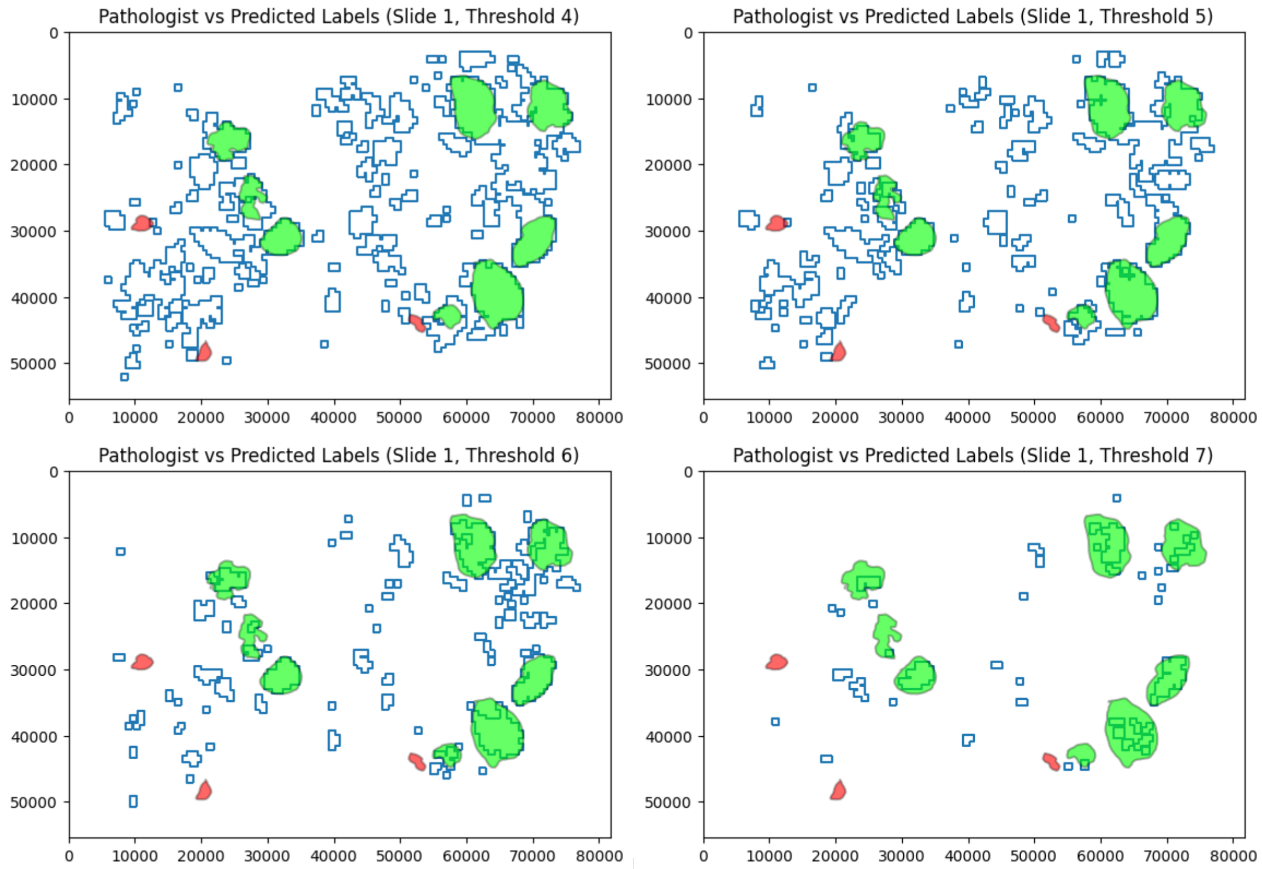
I adapted an existing tumor-annotation model to endometrial biopsy slides using supervised fine-tuning on additional slides annotated by a pathologist. I then refined the prediction threshold to bring the automated tumor regions into closer agreement with those annotations.
Building on earlier research
Earlier work published in Nature Communications identified a p53abn-like subgroup within endometrial cancers classified as NSMP (no specific molecular profile), associated with poorer outcomes. My follow-on contribution focused on adapting the tumor-annotation stage of the image-analysis pipeline from surgical sections to biopsy slides. The model needed to handle the different tissue presentation in biopsy slides. I used additional biopsy images with pathologist-provided annotations for supervised fine-tuning, improving automated tumor annotation on these specimens.
Choosing the prediction threshold
After fine-tuning, I compared predicted tumor regions with the pathologist's annotations across threshold settings. Threshold selection further improved their agreement. The figure illustrates the trade-off: increasing the threshold removes predicted regions outside the annotated tumor, but can also exclude parts of the tumor itself.